Investigating a pleural effusion
The commonest cause for pleural effusion is pneumonia when it's called a parapneumonic effusion. But if a patient comes with pleural effusion with no evidence of an infection, that effusion is considered malignant unless proven otherwise.

The first investigation in any pleural effusion is a chest Xray.
- It will confirm the diagnosis
- There will be aetiological clues like
- Pneumonic patch
- Malignant lesion
- Rib erosions
- Infarctions
Nowadays, an ultrasound scan of the chest is done routinely in all patients with effusions. USS can sometimes visualize consolidations, masses and pleural diseases that might not be apparent on the Xrays. When a malignancy is suspected or the cause is not evident in other investigation modalities, CT becomes the most useful investigation.
The most important investigation in any pleural effusion is the pleural fluid aspiration. You all have to know the technique of doing it. But, now the blind approach is no longer recommended and it always has to be done under ultrasound guidance.
Let's see how to interpret pleural fluid reports.
- First of all, you have to know whether the effusion is an exudate or a transudate.
- For that Lights criteria has to be used.

- Then the cell count can be helpful.
- Neutrophilic effusions - Parapneumonic
- Lymphocytic effusions - Malignant, Tuberculous, lymphoma, Rheumatoid
- Eosinophilia - You might not be questioned on this!
- Mesothelial cells - You might not be questioned on this too!
- Pleural fluid sugar
- Any inflammatory condition will give rise to low sugar. Rheumatoid pleural effusions are known to have very low sugar values.
- pH
- If the pH is less than 7.2, it indicates empyema (Pus in the thorax). That needs IC tube insertion.
- Cytology
- You need to send at least 50ml fluid to the lab.
- Presence of malignant cells indicates a malignant effusion
- Adenosine deaminase (ADA)
- Elevated values are suggestive (Not always) of TB.
- Less commonly used values
- Amylase - Pancreatitis, Esophageal rupture!
- Triglycerides and cholesterol
Remember, in all pleural effusions send samples for gram stain and culture. In Sri Lanka, all need to be sent for AFB, TB PCR, and TB Culture.
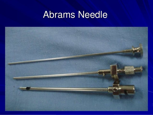
When you suspect TB or malignancy, pleural biopsy is also helpful. It has to be done under USS or CT guidance. The needle (Abrams) is a pet OSCE instrument!
Well, I deliberately avoided blood investigations and other supportive investigations like Mantoux. You can read about them!


Comments
Post a Comment